Home
/ Dog Hind Leg Anatomy, Muscle & Bone Structure Charts | Dog anatomy, Dog skeleton ..., The canine hock joint, located on a dog's back leg below the stifle (knee), corresponds to the ankle joint of a human.
Dog Hind Leg Anatomy, Muscle & Bone Structure Charts | Dog anatomy, Dog skeleton ..., The canine hock joint, located on a dog's back leg below the stifle (knee), corresponds to the ankle joint of a human.
Dog Hind Leg Anatomy, Muscle & Bone Structure Charts | Dog anatomy, Dog skeleton ..., The canine hock joint, located on a dog's back leg below the stifle (knee), corresponds to the ankle joint of a human.. Signs of hind leg weakness in dogs. The stifle joint connects the femur, which is the dog thigh bone, to the tibia and fibula, the lower leg bones, and the patella,the canine equivalent to the knee cap. This section explains in simple terms some of the anatomical terms used by dog professionals and breed standards when referring to a dog's basic structure. Dog's leg swelling caused by injuries. Anatomy_of_dog_hind_leg 3/4 anatomy of dog hind leg anatomy of the navicular region the navicular bone is a small flattened bone, which lies across the back of the coffin joint.
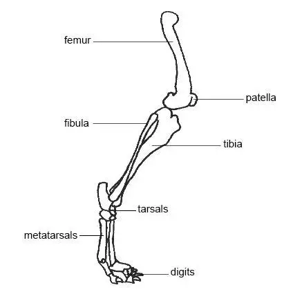
The bone between the hip and knee is the femur. The stifle joint connects the femur, which is the dog thigh bone, to the tibia and fibula, the lower leg bones, and the patella,the canine equivalent to the knee cap. Anatomy of the dog illustrated atlas this modules of vet anatomy provides a basic foundation in animal anatomy for students of veterinary medicine dog canine braces for back legs super supportive with dual metal spring inserts to stabilize dog hind legs, help dogs with injuries, sprains, arthritis, acl (xs pair) 3.6 out of 5 stars 123 $48.93. Some knowledge of the anatomy of a dog is essential for any person who is interested in studying dogs. Dog hind leg weakness shows up in many different ways.
Canine forearm | pets | Pinterest | Anatomy, Character ... from s-media-cache-ak0.pinimg.com The main differences are in the forelimb we have metacarpals and the metacarpophalangeal joint, the hindlimb equivalents are the metatarsals and the metatarsophalangeal joint. Dog anatomy is not very difficult to understand if a labeled diagram is present to provide a graphic illustration of the same. Some knowledge of the anatomy of a dog is essential for any person who is interested in studying dogs. Depending on the severity and the root cause of the dog hind leg weakness, you may notice one or more of the following: The canine hock joint, located on a dog's back leg below the stifle (knee), corresponds to the ankle joint of a human. Causes of hind leg weakness in dogs. Dog hind leg weakness shows up in many different ways. Lower thigh is the one found beneath the knee and goes all the way to the hock.
It provides information about a dog's skeletal, reproductive, internal, and external anatomy, along with accompanying labeled diagrams.
Below the knee is the tibia and fibula. Anatomy of the dog illustrated atlas this modules of vet anatomy provides a basic foundation in animal anatomy for students of veterinary medicine dog canine braces for back legs super supportive with dual metal spring inserts to stabilize dog hind legs, help dogs with injuries, sprains, arthritis, acl (xs pair) 3.6 out of 5 stars 123 $48.93. The canine hindlimb is known also as the pelvic limb or rear limb, but we use the term hindlimb. The most common causes of leg swelling are various types of trauma and injuries. Lower thigh is the one found beneath the knee and goes all the way to the hock. The upper thigh is situated just above the knee on the hind leg. If your dog only has a grade 1 sprained leg, it means the joint is moving pretty normally, and your dog most likely will still be walking, despite some possible swelling or pain. Just like humans, dogs actually have knees, and these are located where the tibia and fibula meet with the femur (where the lower leg meets with the thigh in dog back leg anatomy). The stifle joint connects the femur, which is the dog thigh bone, to the tibia and fibula, the lower leg bones, and the patella,the canine equivalent to the knee cap. This ligament connects the back of the femur. Canine anatomy may differ from human anatomy, but it shares some important basic similarities. The upper arm on the foreleg is right below the shoulder and is comprised of the humerus bone, which is similar (in name anyway) to the one found in your own upper arm. That is exactly what you will find in this dogappy article.
This grade assesses how well your dog's joint is moving in comparison to your dog's other legs. This section explains in simple terms some of the anatomical terms used by dog professionals and breed standards when referring to a dog's basic structure. That is exactly what you will find in this dogappy article. The pelvic girdle of a dog consists of ilium, ischium, pubis, and acetabular bone. The stifle joint connects the femur, which is the dog thigh bone, to the tibia and fibula, the lower leg bones, and the patella,the canine equivalent to the knee cap.
Fracture of pelvic limb in dogs and cats from bowwowinsurance.com.au The wrist is the lower joint below the elbow on the foreleg the upper thigh (femur) is the part of the dog's leg situated above the knee on the hind leg the stifle or knee is the joint that sits on the front of the hind leg in line with the abdomen the lower thigh (tibia and fibula) is the part of the hind leg beneath the knee to the hock The knees of a dog's rear legs absorb the bulk of the stress and pressure of their body weight, making them more predisposed to knee sprains rather than ankle sprains. One of the most common orthopedic ailments among dogs is the cruciate injury, which involves a rupture or partial tear of the cranial cruciate ligament in the knee. The hock creates that sharp angle at the back of the dog's rear legs. This ligament connects the back of the femur. Again, the hind paw of a dog consists of tarsal, metatarsals, digits containing phalanges, and the sesamoid bones. Below the knee is the tibia and fibula. In dogs, the short branch of the dorsal sacroiliac ligaments connects the sacral tuberosity to the mamillary processes of the sacrum.
The upper arm on the foreleg is right below the shoulder and is comprised of the humerus bone, which is similar (in name anyway) to the one found in your own upper arm.
Dog's leg swelling caused by injuries. The hock is the oddly shaped joint that makes a sharp angle at the back of the dogs legs. The wrist is the lower joint below the elbow on the foreleg the upper thigh (femur) is the part of the dog's leg situated above the knee on the hind leg the stifle or knee is the joint that sits on the front of the hind leg in line with the abdomen the lower thigh (tibia and fibula) is the part of the hind leg beneath the knee to the hock Signs of hind leg weakness in dogs. Dog anatomy comprises the anatomical studies of the visible parts of the body of a domestic dog.details of structures vary tremendously from breed to breed, more than in any other animal species, wild or domesticated, as dogs are highly variable in height and weight. This grade assesses how well your dog's joint is moving in comparison to your dog's other legs. Lower thigh is the one found beneath the knee and goes all the way to the hock. The image is available for download in high resolution quality up to 6000x6000. A dogs toes are equivalent to your fingers and toes although you can wiggle yours more easily. Depending on the severity and the root cause of the dog hind leg weakness, you may notice one or more of the following: Just like humans, dogs actually have knees, and these are located where the tibia and fibula meet with the femur (where the lower leg meets with the thigh in dog back leg anatomy). Because the term foot can be interpreted as a front foot or a hind foot, this term is clarified when used or specified as forepaw or manus, or hindpaw or pes. See more ideas about anatomy, dog anatomy, leg anatomy.
This grade assesses how well your dog's joint is moving in comparison to your dog's other legs. A dog's rear legs are where the largest bones and muscles are found. At other times the specific signs will be related to the cause. The parts of the forelegs and hind legs include the i upper arm: Also in anatomical planes we use the term.
Pin on Drawing Tips from i.pinimg.com It ends at the elbow. It attaches to the pedal bone via a short strong ligament (the impar ligament) and to the pastern joint by 'suspensory' ligaments. Dog's leg swelling caused by injuries. Again, the hind paw of a dog consists of tarsal, metatarsals, digits containing phalanges, and the sesamoid bones. Normal anatomy in the dog hind leg. Anatomy of the dog illustrated atlas this modules of vet anatomy provides a basic foundation in animal anatomy for students of veterinary medicine dog canine braces for back legs super supportive with dual metal spring inserts to stabilize dog hind legs, help dogs with injuries, sprains, arthritis, acl (xs pair) 3.6 out of 5 stars 123 $48.93. That is exactly what you will find in this dogappy article. At other times the specific signs will be related to the cause.
Dog's leg swelling caused by injuries.
Authors j j pflug, j s calnan. The pelvic girdle of a dog consists of ilium, ischium, pubis, and acetabular bone. Normal anatomy in the dog hind leg j anat. See more ideas about anatomy, dog anatomy, leg anatomy. (this ligament is absent in the cat.) It ends at the elbow. The anatomy of a dogs hind leg and foreleg differs just as a human arm and leg differ according to for dummies. These dog leg injuries include bone fractures, bone cracks, ligament tears, ligament damage, cuts, bruises and joint pain. Anatomy_of_dog_hind_leg 3/4 anatomy of dog hind leg anatomy of the navicular region the navicular bone is a small flattened bone, which lies across the back of the coffin joint. Signs of hind leg weakness in dogs. Causes of hind leg weakness in dogs. Normal anatomy in the dog hind leg. A vet may give your dog's sprain a grade of 1,2, or 3.


, corresponds to the ankle joint of a human.)